RESEARCH • Lung Ultrasound in a Tertiary Intensive Care Unit Population: A Diagnostic Accuracy Study
Source: Crit Care (2021) 25:339
BACKGROUND
Etiology of respiratory failure in ICU patients include cardiogenic pulmonary edema (CPE), acute respiratory distress syndrome (ARDS), atelectasis, and pneumonia . Chest X-ray and thoracic computed tomography (CT) are still the most common diagnostic modalities of ICU patients with respiratory failure.
Previous studies comparing lung ultrasound to thoracic CT findings in ICU patients suffered from multiple methodologic limitations. Most studies excluded patients with multiple diagnoses, were inadequately blinded, or did not include anterior consolidations and/or pneumothorax.
Therefore, the authors undertook this study to evaluate the diagnostic accuracy of a 6-zone lung ultrasound protocol in ICU patients, with thoracic CT as reference standard, using adequate blinding and including patients with multiple respiratory conditions. Secondary aims were:
- To evaluate the diagnostic accuracy of an extended 12-zone lung ultrasound protocol
- To correlate lung ultrasound patterns with respiratory conditions in a tertiary ICU patient population
METHODS
Study design
This was a single-center, prospective, observational diagnostic accuracy study conducted at a tertiary intensive care unit (ICU).
Study population
Patients older than 18 years who admitted to the ICU who underwent thoracic CT for any clinical indication. Those included mechanically and nonmechanically ventilated patients. Patients were excluded if thoracic CT and lung ultrasound examination were more than 8 h apart.
The 17 lung ultrasound examiners were blinded for any CT findings. Patient characteristics collected from the electronic patient record were sex, age, body mass index, past medical history, sequential organ failure assessment (SOFA) score at day of CT-scan, reason for ICU admission, indication for CT-scan and respiratory conditions.
Index test
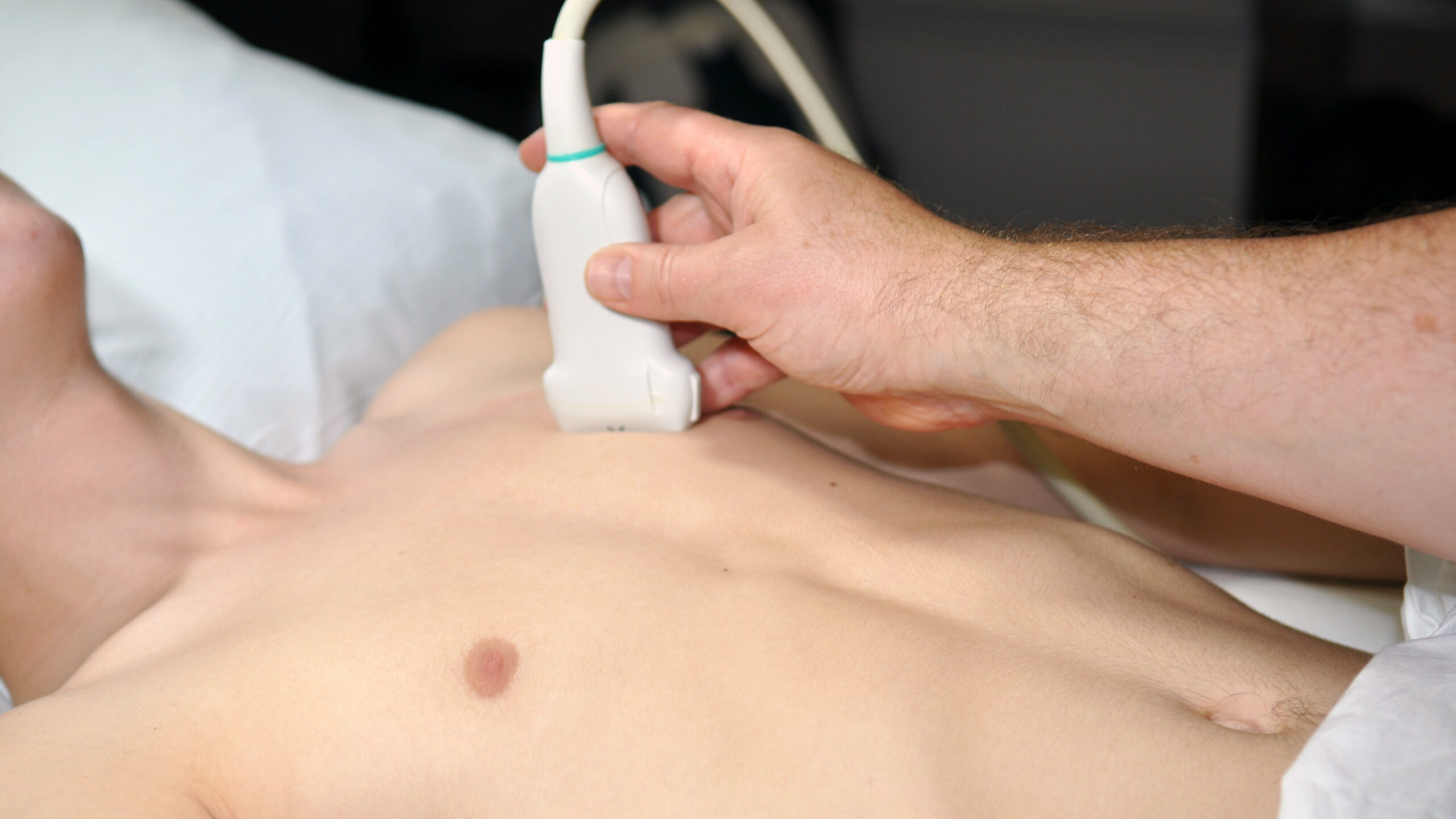
Index test consisted of a 6-zone lung ultrasound examination according to the Bedside Lung Ultrasound in Emergency (BLUE)-protocol. The upper and lower BLUE-points were used to evaluate the anterior lung region, and the posterolateral alveolar and/or pleural syndrome (PLAPS)-point was used to evaluate the posterior lung region. A linear high-frequency transducer was used to evaluate the anterior lung region. For the PLAPS-point a low-frequency, convex, phased array transducer was used. During ultrasound evaluation with the linear transducer, artifact eliminating software was turned off.
Each hemithorax was classified according to the following lung patterns:
- Anterior consolidation: C-profile or B-profile without lung sliding (B’-profile) at the upper or lower BLUE-point
- Posterior consolidation: hypoechoic image restricted by an irregular border or a tissue-like pattern at the PLAPS-point
- Interstitial syndrome: B-profile with lung sliding at upper or lower BLUE-point. This classification requires the absence of an anterior consolidation
- Pneumothorax: A-profile without lung sliding at upper or lower BLUE-point
- Pleural effusion: anechoic or hypoechoic area between parietal and visceral pleura at any point
To potentially increase the sensitivity of lung ultrasound, a more elaborate 12-zone protocol was used in a subgroup of patients. In this group, besides the BLUEpoints and PLAPS-point, the anterior axillary line and posterior axillary line were divided into a superior and inferior half.
Reference test
Thoracic CT was the reference test for this study. CT images were assessed and reported by a radiologist blinded for the results of lung ultrasound examination. All images were evaluated using a mediastinal window and a lung window setting. Lung findings reported by the radiologist were adjudicated to their respective location (anterior or posterior).
Outcomes
Primary outcome was the diagnostic accuracy of lung ultrasound to detect consolidation (anterior or posterior consolidation), interstitial syndrome, pneumothorax and pleural effusion as compared to thoracic CT.
secondary outcomes included the diagnostic accuracy of an extended, 12-zone lung ultrasound protocol and correlation of lung ultrasound patterns with respiratory conditions.
RESULTS
87 patients were included in the primary analysis. For secondary analysis (patients who received a 12-zone lung ultrasound examination) 18 patients were included.
- Prevalence of consolidations was 0.50 (156/311). Estimated sensitivity and specificity were 0.76 and 0.92, respectively.
- Prevalence of interstitial syndrome was 0.51 (80/158). Estimated sensitivity and specificity were 0.60 and 0.69, respectively.
- Prevalence of pneumothorax was 0.11 (17/158). Estimated sensitivity and specificity were 0.59 and 0.97, respectively.
In total there were 147 respiratory conditions in 79 patients. The majority (49.4%, 39/79) had two respiratory conditions. The most prevalent lung pathology was atelectasis, followed by pneumonia. The least prevalent lung pathology was lung infarction
CONCLUSIONS
The authors concluded that lung ultrasound is an adequate diagnostic modality in a tertiary ICU population to detect consolidation, clinically significant pneumothorax and pleural effusion and is less suitable to detect interstitial syndrome.
They also concluded that a 12-zone lung ultrasound protocol had no added benefits over the 6-zone lung ultrasound protocol.
Finally, they commented that «one should be careful not to interpret lung ultrasound findings in deterministic fashion as most patients have multiple respiratory conditions associated with (partially) similar lung ultrasound patterns.»
 Español
 English
