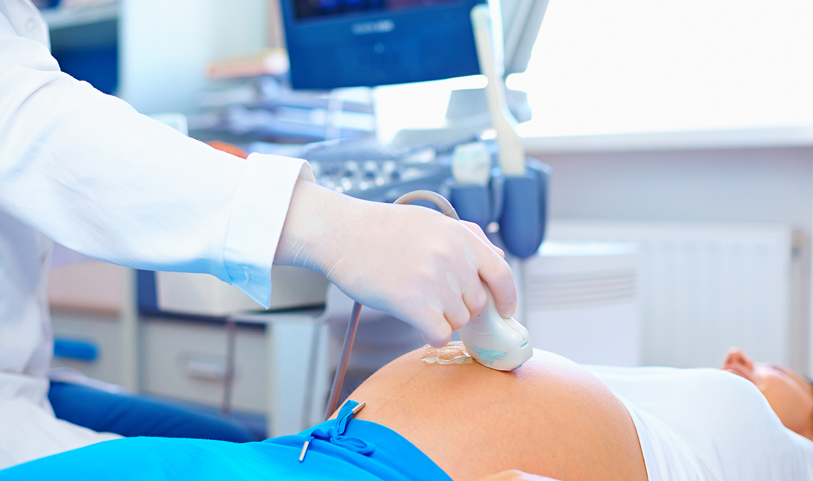
Point-of-care ultrasound in pregnancy: gastric, airway, neuraxial, cardiorespiratory.
Source: Curr Opin Anesthesiol 2020, April 18
INTRODUCTION
This is a review article that looks into the use of PoCUS in the anesthetic management of the obstetric patient. The article reviews the use of ultrasound for assessment of aspiration risk, airway management, placement of neuraxial blocks and the diagnosis and follow-up of cardiorespiratory dysfunction.
GASTRIC ULTRASOUND
Pregnant patients at term are considered high-risk for aspiration because of anatomical and physiological changes and the urgency of interventions. In addition, they have a higher chance of a difficult airway, which makes an aspiration event potentially catastrophic. Gastric PoCUS can be used to assess aspiration risk and potentially avoid aspiration.
The antrum is evaluated for qualitative (empty, solid, clear fluid, thick fluid) and quantitative nature (total gastric fluid volume estimation). This approach may permit to assess aspiration risk and to tailor the anesthetic technique for each patient. The antrum is identified in both the supine and right-lateral decubitus (RLD) position with a low-frequency transducer in the epigastrium. The empty antrum is flat with a bull’s eye appearance. Solid food can be appreciated in a dilated antrum as hyperechoic content with ring-down artifacts. Clear fluids present as hypoechoic content.
The amount of clear fluids can be estimated by measuring the antral cross-sectional area (CSA) in the RLD and the use of a validated mathematical model. Among all, the Perlas model is probably the most widely used: GV (ml) = 27 + (14.6 x right-lat CSA) ! (1.28 x age). The Perlas grading system offers a simple and alternative identification of low-volume versus high-volume states based on the qualitative assessment of the antrum only. It is defined as grade 0 when it appears empty in both positions. It is classified as grade 1 when fluid is present in the RLD only, which correlates with low gastric volumes. A grade 2 antrum (fluid in both positions) correlates with a high aspiration risk.
AIRWAY ULTRASOUND
Physiological changes during pregnancy such as obesity, enlarged breasts and fluid retention that causes soft tissue mucosal airway edema lead to higher incidences of difficult airway when compared to non-pregnant patients (at least 10 times higher than in the general surgical population). The failed intubation rate in this patient population has remained unchanged between 1970 and 2015.
Accurate airway assessment, which is the most important step in airway management, must be performed before induction of general anesthesia. Airway assessment tests – evaluation of Mallampati classification, thyromental distance, interincisor distance and BMI – have low sensitivity and specificity with limited and unreliable predictive value.
Airway ultrasound evaluation may provide better assessment of the epiglottis, vocal cords and membranes, and can confirm correct tracheal tube placement. In addition, it increases the rate of accurate identification of the cricothyroid membrane compared with palpation. This may be important in emergencies, since accurate knowledge of the airway depth before emergency may improve the cricothyroidotomy success rate.
ULTRASOUND FOR NEUROAXIAL ANESTHESIA
Although neuraxial anesthesia is highly recommended for the obstetric patient, it can be very challenging in this pa+ent population. Locating the midline for the identification of the inter-vertebral space for needle entry is often very challenging. The landmark technique uses palpation of the vertebral spinous processes for identification of the midline and planning of the procedure. However, in the parturient patient the identification of the inter-vertebral space may be near impossible due to the obliteration by large amount of adipose tissue.
Ultrasound imaging is used to facilitate needle placement by identifying the midline of the spine and the intervertebral level and measuring epidural space depth. Compared with the traditional landmark technique, preprocedural ultrasound reduces the number of punctures, decreases the risk of failed epidural analgesia and is associated with higher pa+ent satisfaction. This is probably more beneficial in patients with difficult spinal anatomy or high BMI, showing that even experienced anesthesiologists achieve higher first-pass rates and shorter needling +me when using ultrasound.
 English
 Español

