Clinical Research: Tissue Doppler Imaging of the Diaphragm in Healthy Subjects and Critically Ill Patients
Source: American Journal of Respiratory and Critical Care Medicine. Published online: July 2, 2020
INTRODUCTION:
In recent years, ultrasonography has been used to evaluate diaphragmatic motion characteristics such as excursion and thickening, with derived measurements being shown to relate to outcome of weaning from mechanical ventilation.
Tissue Doppler Imaging (TDI) is an echocardiographic technique that uses the Doppler principle of detecting the frequency shift of ultrasound signals reflected by moving objects to measure the high amplitude, low velocity signal of cardiac tissue motion.
This study was designed as a two-stage prospective observational study. First, the authors evaluated the waveform pattern of diaphragmatic TDI, to measure the velocities of diaphragmatic contraction and relaxation in healthy individuals, report the normal values and assess the reproducibility of the method. In the second stage, the authors recorded the diaphragmatic TDI pattern and values in intubated ICU patients during a spontaneous breathing trial, comparing weaning success and weaning failure cases.
In a subgroup of patients, the authors also correlated TDI measurements to parameters derived from the corresponding transdiaphragmatic pressure (Pdi) waveforms, aiming at a better understanding of the clinical significance of the TDI values and their derived parameters.
METHOD
In the first stage, TDI was performed on twenty healthy volunteers (ten men and ten women, aged between 25-48 years), where diaphragmatic TDI waveform was assessed. This allowed the authors to describe the range of normal values of the diaphragmatic motion velocities during inspiration and expiration, and to evaluate the reproducibility of the method. A total of over 150 breaths were recorded, accounting for about 8-10 breaths per participant.
In the second stage, consecutive ICU patients who were mechanically ventilated for more than 48 hours were screened for participation in the study. Patients were excluded if they were younger than 18 years of age, had neuromuscular diseases, flail chest or multiple rib fractures, had chest tubes in place, were morbidly obese, had spinal cord injuries, or if TDI signal could not be obtained from the right hemidiaphragm.
The sonographic examination was performed at the end of a 30 minutes T-piece weaning trial or immediately before reconnection to the ventilator in the case of weaning failure occurring before.
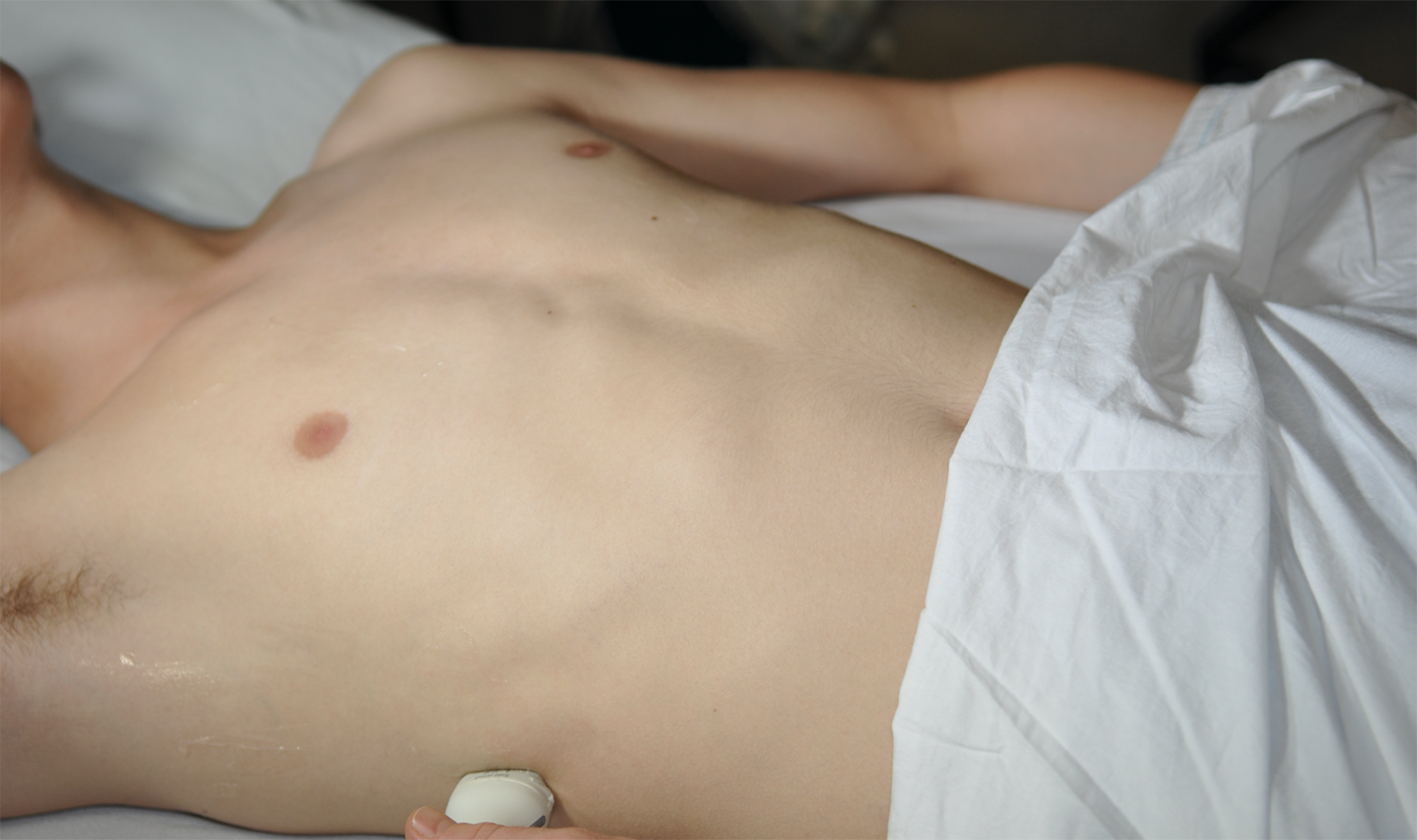
Since TDI is unable to discriminate passive from active motion, patients were assessed during spontaneous breathing because TDI assesses only the absolute tissue velocity. Since the acoustic window on the left are limited, all ultrasound measurements were performed on the right hemi-diaphragm. TDI images were obtained with a phased array 2-4 MHz probe («cardiac probe»), which was placed in the subcostal position between the mid-clavicular and anterior axillary lines, with the patients being examined while breathing spontaneously laying on a bed with the back elevated at 30°.
RESULTS
Stage 1 – control group
A total 168 different breaths from the control group of 20 healthy volunteers were recorded (about 810 breaths per participant). Excellent intra- and inter-observer reproducibility was found for all variables.
Stage 2 – study population
A total of 116 consecutive ICU patients were enrolled in the study. Sixty-seven patients (57.8%) succeeded the weaning trial. Diaphragmatic assessments in these patients included measurements of diaphragmatic displacement and thickening, followed by TDI measurements. When TDI-derived values from ICU patients were compared to those recorded in healthy controls, peak contraction and peak relaxation velocities did not differ between weaning success patients and healthy volunteers.
Transdiaphragmatic pressure (Pdi) Measurements: All three Pdi-derived parameters – Peak Pdi (peak value of Pdi measured in cmH2O), PTPdi (diaphragmatic pressure-time product measured cmH2Oꞏsec), and Pdi-MRR (the diaphragmatic maximal relaxation rate) – were significantly lower in patients who succeeded the weaning trial (n=8) than in weaning-failure patients (n=10).
DISCUSSION
The authors comment on the fact that no previous information exists about the diaphragmatic TDI pattern and range of normal values in normal individuals or in ICU patients. This study established the TDI pattern of diaphragmatic contraction and relaxation, along with their peak corresponding values, the inspiratory velocity-time integral and the TDI-derived relaxation rate in normal individuals as well as in patients who successfully were able to wean off the ventilator and those who did not. The authors found that patients who failed to wean off the ventilator exhibited significantly higher peak contraction and relaxation velocities and higher relaxation rate compared to both healthy participants and patients who were able to successfully wean off the ventilator.
Pdi measurement assesses diaphragmatic contractility and represents and index of diaphragmatic contractile performance. However, its measurement is limited by technical challenges. In addition, there is lack of precise reference values in critically ill patients. Therefore, the widespread clinical application of the method is restricted.
The study correlated peak Pdi with TDI peak contraction velocity with high correlation coefficients, suggesting a potential application of this non-invasively acquired parameter to quantify diaphragmatic performance.
 Español
 English
